Microscopy and cellular imaging

Leader: Mathieu ERHARDT

Description

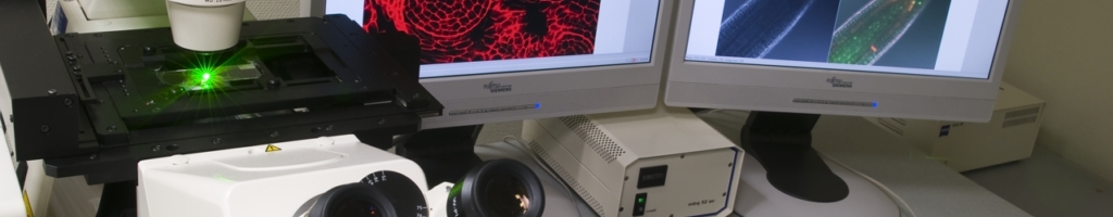
The microscopy facility was the first technology platform established in the IBMP in 1998. It’s scientific programme aims at understanding the expression of plant or animal genes over space and time at various levels. Microorganisms or biomaterials studied by partner research units are other topics of interest. Our facility follows official guidelines for « Plates-Formes Technologiques du Vivant » and has received RIO 2001, 2004 and 2006 labels. It’s part of the larger Strasbourg Esplanade Cell Imaging Facility that allows sharing devices and knowledge from 7 research units from CNRS, INSERM, UNISTRA and INRA.

Missions

  • Assist research from IBMP and partner research units
  • Develop and implement new imaging technologies
  • Give appropriate one-to-one or group training to research staff
  • Get involved in microscopy education and science popularization
  • Make sure our devices are up and running

Expertises

Electron Microscopy and histology

Manager : Mathieu ERHARDT

Provides expertise, development and training for the following technologies:

  • TEM, SEM, serial block face imaging
  • Histology, cytology, in situ hybridization

Optical microscopy and bioimage analysis

Manager : Jérôme MUTTERER

Provides expertise, development and training for the following technologies:

  • Brightfield , fluorescence, confocal microscopy, fluorescence lifetime imaging
  • Image Processing

Members

Recent publications

  • SCHIAFFINI M., CHICOIS C., POUCLET A., CHARTIER T., UBRIG E., GOBERT A., ZUBER H., MUTTERER J., CHICHER J., KUHN L., HAMMANN P., GAGLIARDI D. and GARCIA D.

    A NYN domain protein directly interacts with DECAPPING1 and is required for phyllotactic pattern

    Plant Physiology, 188(2):1174–1188, 2022. | DOI : 10.1093/plphys/kiab529DOI logo

  • HUANG C., MUTTERER J. and HEINLEIN M.

    In vivo aniline blue staining and semi-automated quantification of callose deposition at plasmodesmata

    Methods in Molecular Biology, 2457:151-165, 2022. | DOI : 10.1007/978-1-0716-2132-5_9DOI logo

  • CHEVIGNY N., LOTFI F., LE BLEVENEC A., NADIRAS C., FERTET A., BICHARA M., ERHARDT M., DIETRICH A., RAYNAUD C. and GUALBERTO J.M.

    RADA-dependent branch migration has a predominant role in plant mitochondria and its defect leads to mtDNA instability and cell cycle arrest

    PLoS Genetics, e1010202, 2022. | DOI : 10.1371/journal.pgen.1010202DOI logo

  • JOHANN TO BERENS P., SCHIVRE G., THEUNE M., PETER J., SALL S.O., MUTTERER J., BARNECHE F., BOURBOUSSE C. and MOLINIER J.

    Advanced image analysis methods for automated segmentation of subnuclear chromatin domains

    Epigenomes, 6(4):34-40, 2022. | DOI : 10.3390/epigenomes6040034DOI logo

  • CHEVALIER Q., GALLÉ J.B., WASSER N., MAZAN V., VILLETTE C., MUTTERER J., ELUSTONDO M.M., GIRARD N., ELHABIRI M., SCHALLER H., HEMMERLIN A. and VONTHRON-SENECHEAU C.

    Unravelling the Puzzle of Anthranoid Metabolism in Living Plant Cells Using Spectral Imaging Coupled to Mass Spectrometry

    Metabolites, 11:571, 2021. | DOI : 10.3390/metabo11090571DOI logo